What Is 4D Ultrasound?
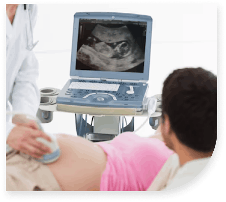
3D/4D ultrasound is an ultrasound technique used during pregnancy to produce lifelike images of the fetus. With this latest Ultrasound technology, a fourth dimension is added to a normal 2D sonography image resulting in a moving real-time image of your unborn child.
The Criteria To Be Eligible For a 4D Scan
3D and 4D ultrasound scans should be regarded as additional scans and are not part of your routine antenatal care. However, 3D and 4D scans provide more information about a known abnormality and are regularly used as part of our anomaly scan whenever necessary. It helps to closely examine suspected fetal anomalies, such as cleft lips or spina bifida. 4D scanning can also be useful to look at the heart and other internal organs. Because these scans can show more details from different angles, this can help doctors in planning the treatment after birth. In addition, our state-of-the-art ultrasound equipment shows how the baby “behaves” in the womb and what your baby looks like before being born.
When Is The Best Time To Have The 3D/4D Scan?
For optimal 3D and 4D scan images, the best time to arrange a visit is between 24 and 28 weeks (after you’ve had your fetal anomaly scan at 18-22 weeks). Women with twins wishing to attend for a 3D and 4D baby scan should visit at around 25 weeks, as twins have less space.
Is 3D/4D Ultrasound Scanning Safe?
There has been no evidence to suggest that Ultrasound scanning presents any risk to the mother and baby. This is being monitored on an ongoing basis by experts worldwide. The Ultrasound output levels of our 3D and 4D scanning equipment, GE Voluson machine are no more than the traditional 2D scanning equipment used. While there is no hard evidence of any harmful effects of 3D/4D ultrasound, its use in non-medical situations should not be undertaken

